


SARS-CoV-2 Identification by TEM
SARS-CoV-2 Identification by TEM
A Powerful Tool in Viral Visualization and Structural Confirmation
The emergence of SARS-CoV-2 during the global pandemic led to an urgent need for rapid and accurate virus identification. While molecular techniques like RT-PCR became the gold standard for diagnosis, Transmission Electron Microscopy (TEM) played a crucial role in visualizing and confirming the structural identity of the virus.
🦠 What is SARS-CoV-2?
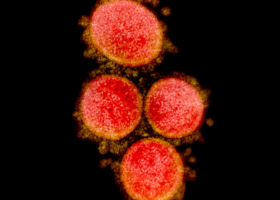
SARS-CoV-2 is the causative agent of COVID-19. It belongs to the coronavirus family and is characterized by its crown-like spike proteins visible under high-resolution microscopy.
🔬 What is Transmission Electron Microscopy (TEM)?
Transmission Electron Microscopy is an advanced imaging technique that uses a beam of electrons to visualize structures at the nanometer scale. Because viruses are extremely small (approximately 60–140 nm in diameter), TEM is particularly effective for observing their morphology.
📌 Why Use TEM for SARS-CoV-2 Identification?
Although molecular diagnostics detect viral RNA, TEM allows:
Direct visualization of viral particles
Confirmation of viral morphology
Structural analysis of spike proteins
Observation of virus-host cell interactions
Validation of viral culture results
TEM was especially important during the early outbreak stages when confirming the novel virus structure.
🧪 Sample Preparation for TEM
Accurate identification requires meticulous preparation:
Sample Collection – Nasopharyngeal swabs, bronchoalveolar lavage fluid, or cultured infected cells.
Fixation – Using glutaraldehyde to preserve viral structure.
Embedding – Resin embedding for ultrathin sectioning.
Ultrathin Sectioning – 50–100 nm slices using an ultramicrotome.
Staining – Heavy metal stains such as uranyl acetate and lead citrate enhance contrast.
🔎 Morphological Features of SARS-CoV-2 Under TEM
When observed under TEM, SARS-CoV-2 shows:
- Spherical to pleomorphic particles
- Diameter of 60–140 nm
- Distinct spike (S) glycoproteins forming a corona-like appearance
- Electron-dense nucleocapsid core
- Localization within cytoplasmic vesicles of infected cells
- These features differentiate it from other respiratory viruses.
⚠️ Challenges in TEM Identification
- Despite its strengths, TEM has limitations:
- Requires specialized infrastructure and expertise
- Lower sensitivity compared to molecular tests
- Potential misinterpretation with similar cellular structures
- Time-consuming sample preparation
Therefore, TEM is generally used as a confirmatory or research tool rather than a routine diagnostic method.
🌍 Role of TEM in the COVID-19 Pandemic
During the initial outbreak in Wuhan, TEM helped scientists visualize the novel coronavirus from patient samples, supporting early structural characterization. It also contributed to understanding viral assembly, replication, and vaccine research.
🧬 Comparison: TEM vs Molecular Methods
| Feature | TEM | RT-PCR |
|---|---|---|
| Detects | Whole viral particles | Viral RNA |
| Sensitivity | Moderate | Very High |
| Speed | Slower | Rapid |
| Use | Research & confirmation | Clinical diagnosis |
📚 Conclusion
Transmission Electron Microscopy remains a vital tool in virology research. While it may not replace molecular diagnostics, it provides unparalleled structural insights into pathogens like SARS-CoV-2. The ability to directly observe viral particles enhances our understanding of virus morphology, replication, and interaction with host cells.
For researchers, microbiologists, and nanotechnology professionals, TEM continues to be an indispensable technique in advanced viral identification and characterization.
Comments
Latest TrendFathima Shereen M
20 May 2026Interesting read! the article explained the visualization of coronavirus particles through TEM and their nanoscale structure.
S Girisha
20 May 2026Very informative article on the role of TEM in SARS-CoV-2 identification. The explanation of sample preparation and viral morphology was clear and easy to understand. It shows how advanced imaging techniques contribute significantly to virology research and structural analysis.
Udayashree M.R.V.
10 Apr 2026Thankyou so much For the clear explanation about what is TEM , and their identification , especially sample preparation - how To prepare samples step by step and their challenges and comparisons on tem and rtpcr. Its very useful and also i need to know more about ftir - how it works , sample preparation etc.....
SUPREEM MANDAL
10 Apr 2026Thankyou so much.
Dr. BALARAM SARKAR
10 Apr 2026to study the ultrastructure of beneficial microorganisms, complex interactions between microbes, plants and carrier materials
MOHD RAZA
10 Apr 2026TEM and FTIR play and important role in structural analysis as well as morphology of virus for study further
ELAKKIYAN M
09 Mar 2026The TEM IS related my project work thanks for your explanation
Rishika Bavishi
08 Mar 2026A clear explanation of the role of TEM in viral visualization and research. Structural insights like these are crucial for advancing virology and vaccine development.
kanika chauhan
08 Mar 2026Great explanation of how TEM contributes to advanced viral research and characterization.
USHASI MUKHERJEE
04 Mar 2026This was very insightful. It clearly explains how TEM helps visualize SARS-CoV-2 and supports molecular diagnostic methods in virology research.